Early detection of gastric cancer significantly increases treatment success rates. However, because stomach cancer often progresses silently, catching it in its early stages requires advanced diagnostic tools. Patients undergoing upper endoscopy in Baltimore benefit from a procedure that allows direct visualization of the upper gastrointestinal tract, enabling clinicians to identify abnormalities that other methods might miss. Endoscopy serves as both a diagnostic and biopsy platform, offering accuracy, speed, and minimal invasiveness.
Understanding the Endoscopic Procedure
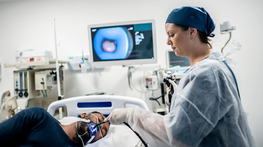
Upper endoscopy, or esophagogastroduodenoscopy (EGD), involves inserting a flexible tube with a high-definition camera through the mouth into the esophagus, stomach, and upper part of the small intestine. This real-time visual inspection enables physicians to detect tissue irregularities, ulcers, inflammation, or suspicious lesions.
Biopsy and Tissue Sampling
When abnormalities are observed, small instruments passed through the endoscope can collect tissue samples without the need for open surgery. These biopsies are then sent for histological analysis to determine if the cells are cancerous, pre-cancerous, or benign. This capability makes endoscopy not just a visual tool, but a critical instrument for early cancer diagnosis.
Recognizing Early Signs
Stomach cancer can present as subtle changes, such as mucosal irregularities, discoloration, or small nodules, that are often missed by imaging alone. Endoscopy offers the advantage of detecting these fine visual clues with clarity. Additionally, advanced endoscopic techniques, such as narrow band imaging (NBI), enhance visualization of blood vessels and tissue structure, improving the ability to differentiate between malignant and non-malignant growths.
For individuals with a family history of gastric cancer, chronic Helicobacter pylori infection, or unexplained gastrointestinal symptoms, early screening via endoscopy is often advised as part of a preventive care plan.
Beyond Detection: Monitoring and Surveillance
Endoscopy is also essential for ongoing surveillance in patients with known risk factors or those undergoing treatment for early-stage gastric cancer. It allows healthcare providers to monitor tissue response to therapy and assess for recurrence or progression. This proactive approach ensures that intervention can be adjusted promptly based on real-time findings.
Patients are often advised to undergo repeat endoscopic evaluations depending on their risk profile and previous findings, making it a cornerstone of both detection and ongoing disease management.
Understanding what the upper endoscopy procedure involves equips patients with the clarity they need to approach this diagnostic step with confidence. Familiarity with the process reduces anxiety and improves cooperation, leading to more accurate results.
Minimally Invasive, Highly Effective
Unlike exploratory surgeries of the past, modern endoscopy is minimally invasive and typically performed under conscious sedation. The procedure usually lasts no more than 20 to 30 minutes, with minimal downtime or recovery required. It offers clinicians the ability to see, sample, and even treat certain findings in a single session, streamlining care and improving efficiency.
Conclusion
Endoscopy stands as one of the most effective tools in the early detection of stomach cancer. With its ability to provide high-resolution imagery and real-time biopsies, it bridges the gap between suspicion and diagnosis.
Professional use of upper endoscopy not only detects abnormalities at a stage when they are most treatable but also supports long-term monitoring and personalized care strategies. For patients and clinicians alike, it remains a critical ally in the fight against gastric cancer, delivering clarity, precision, and early intervention where it matters most.